-
Buyer
Register as a Foriegn Buyer
-
Seller
Log In Register as a Seller
Buyer
Seller


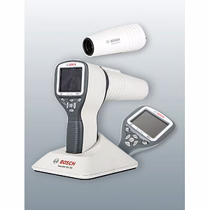
ITC-HSN: 90185090
Min Order Qty 1 Unit
Accept Small Orders
Product not available
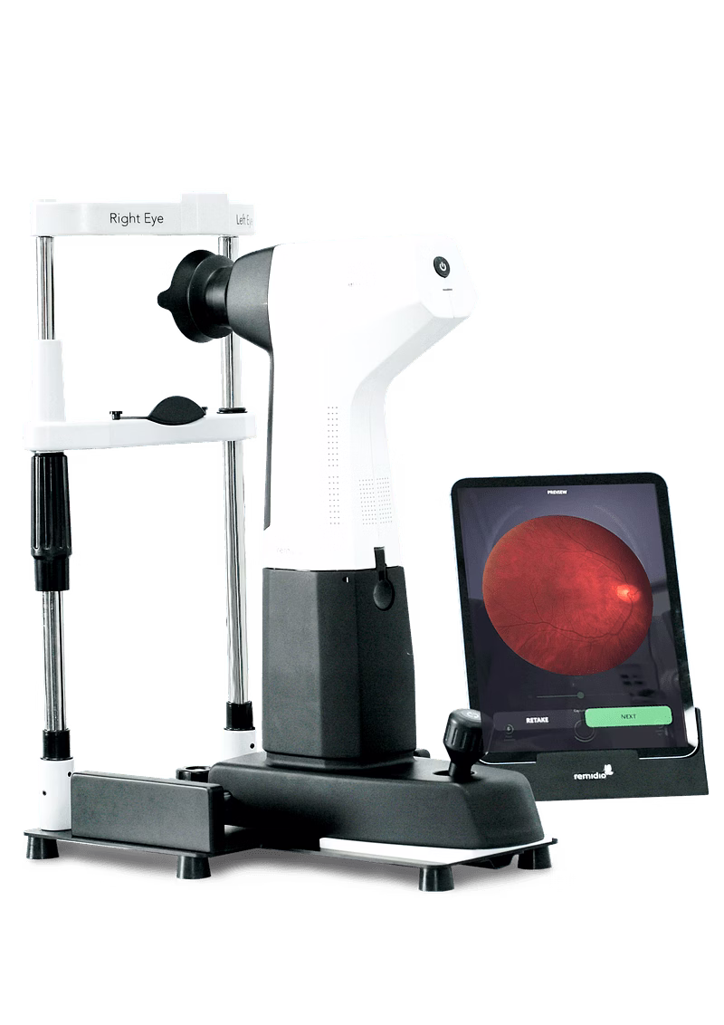
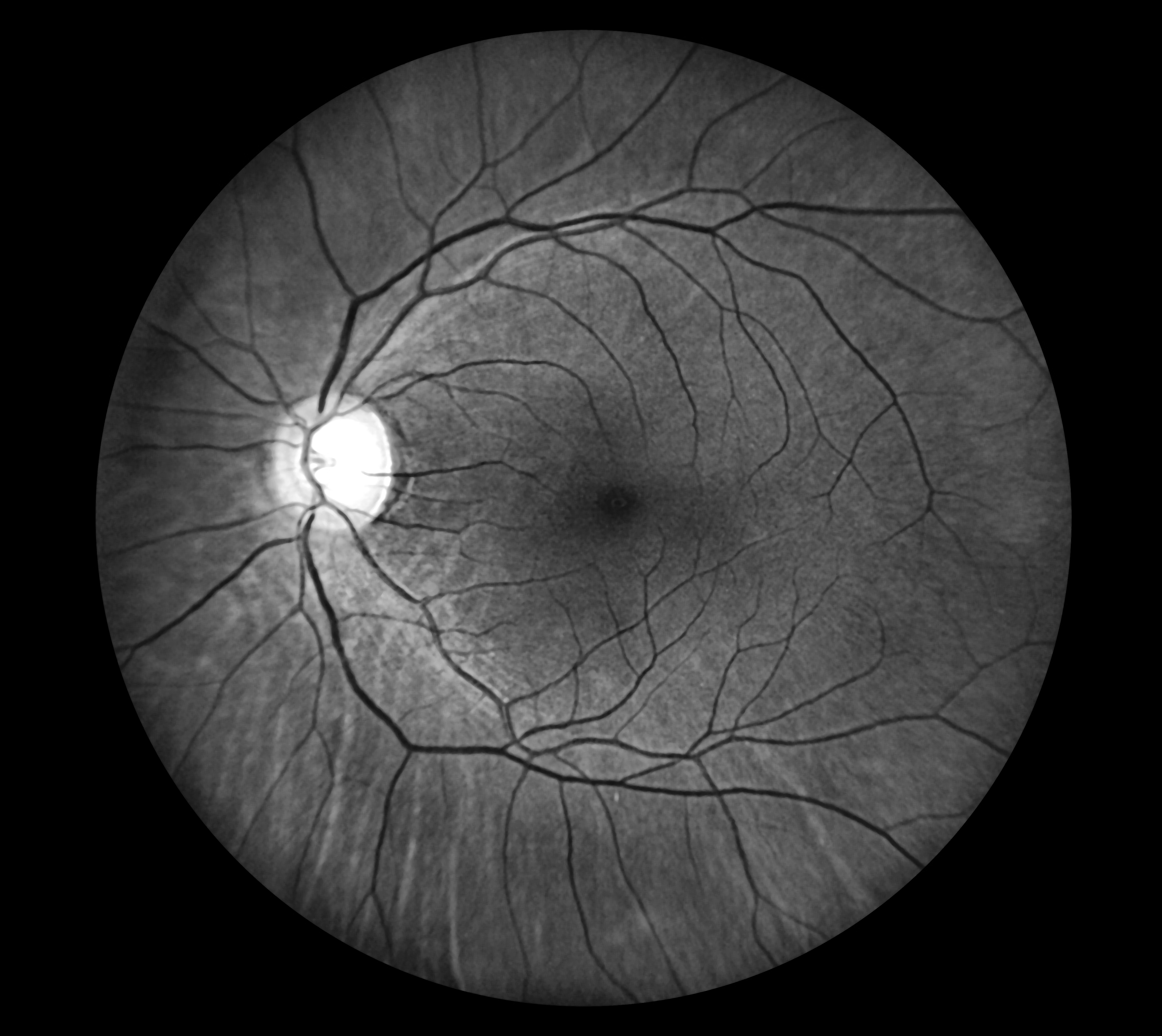
Powered by a 20 MP image sensor and a patented optical design
Wider Field enabling detection of peripheral disease
7-field ETDRS recommends a 75-degree FOV for early detection of Diabetic Retinopathy. With just two clicks, Pristine 5.0 offers upto 87° X 58° FOV*# and up to 87° X 116° FOV*# with four clicks.
*Visual Angle
#Horizontal x Vertical
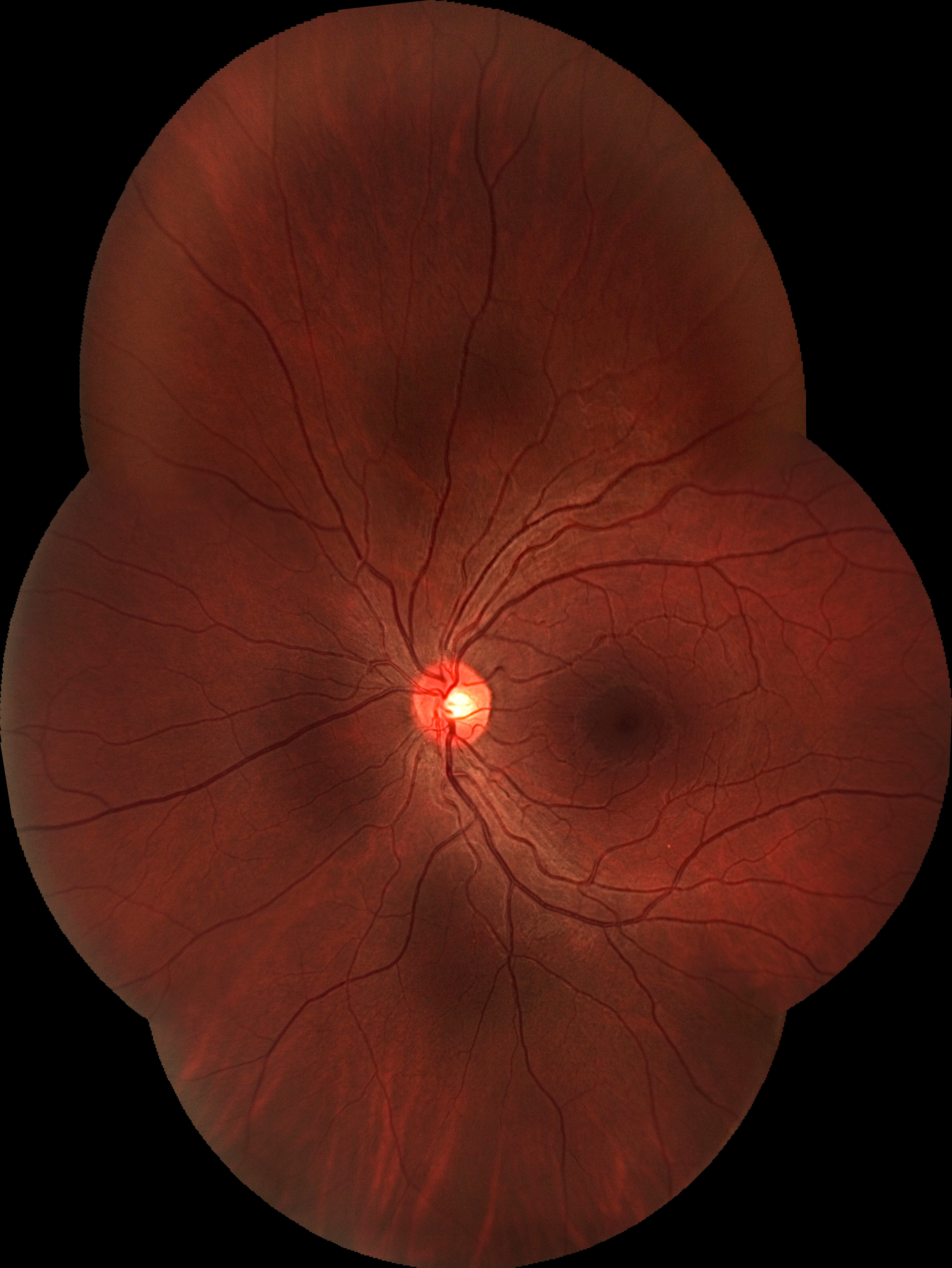
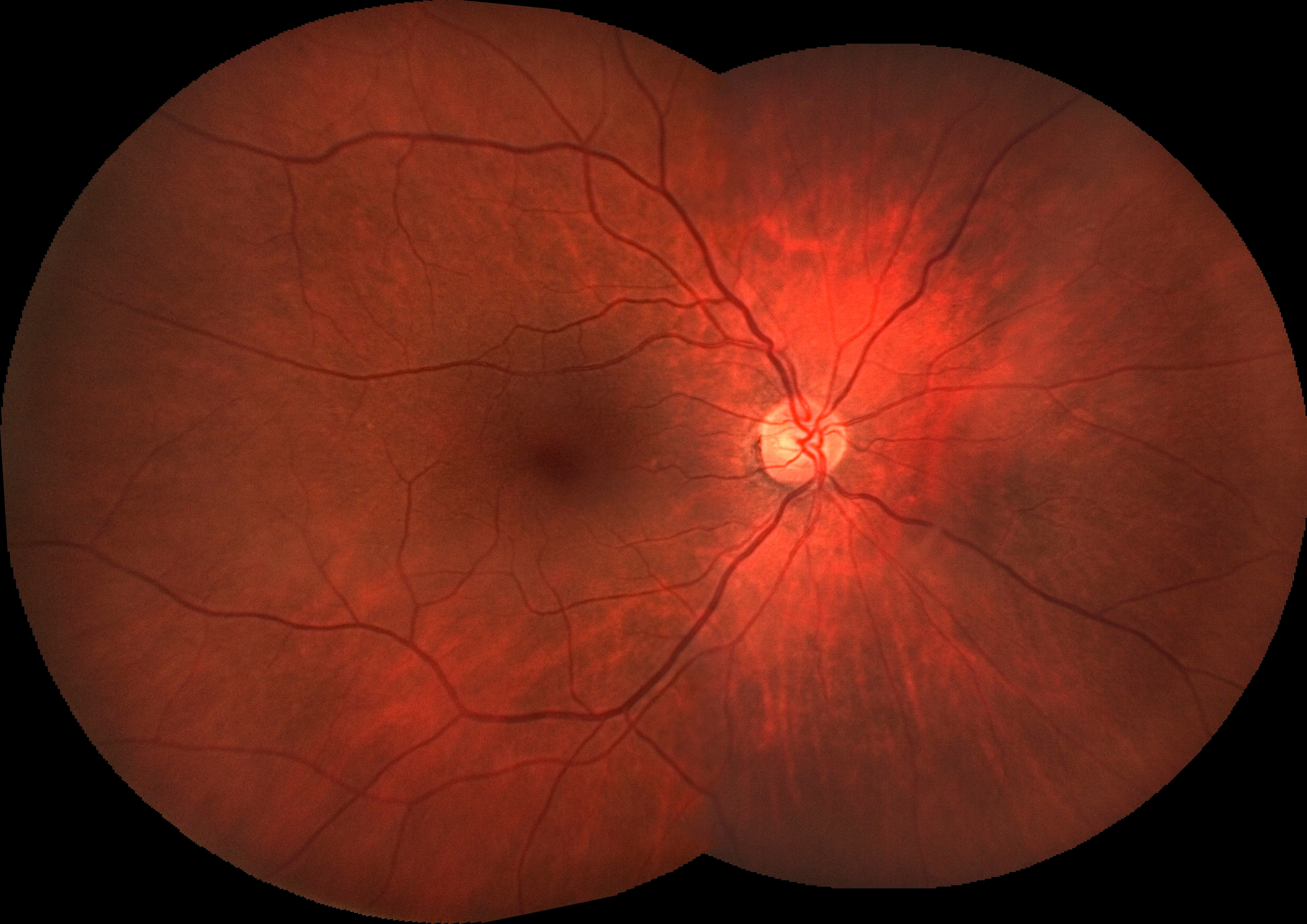
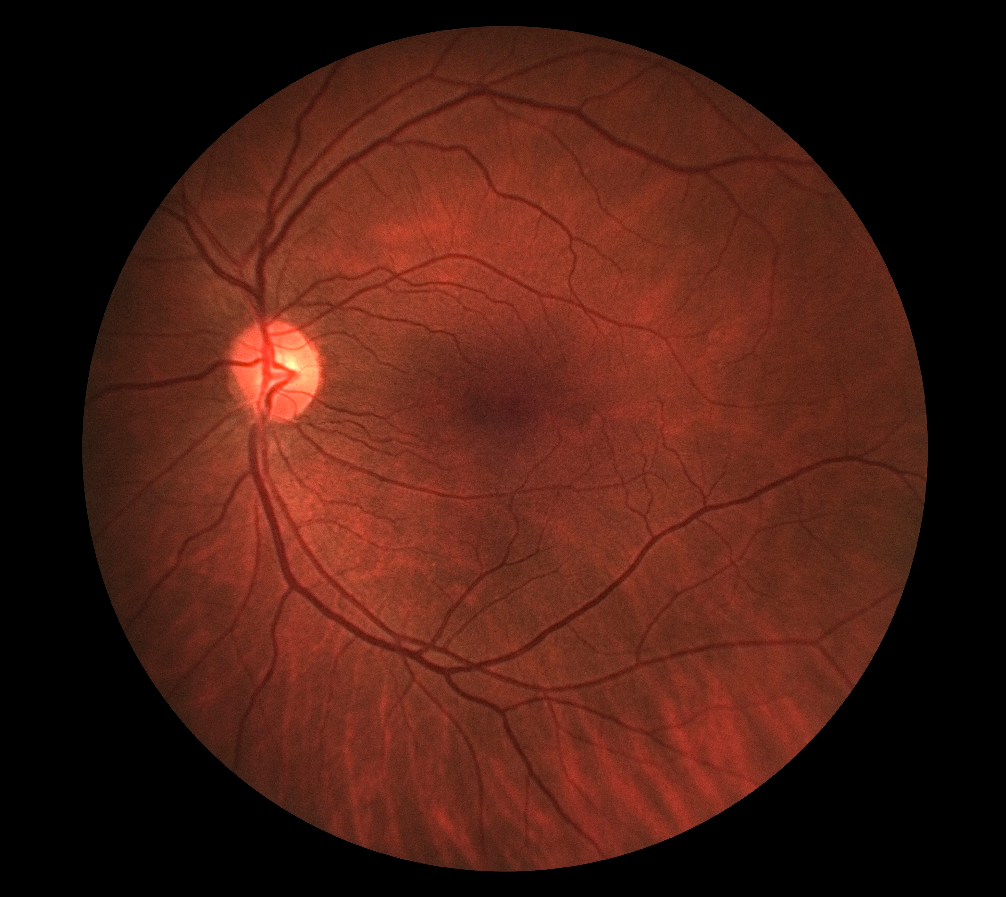
Precision in Clinical Decision
Experience everyday precision with sharp detailing in images that identifies even hard-to-detect pathologies and tertiary blood vessels.
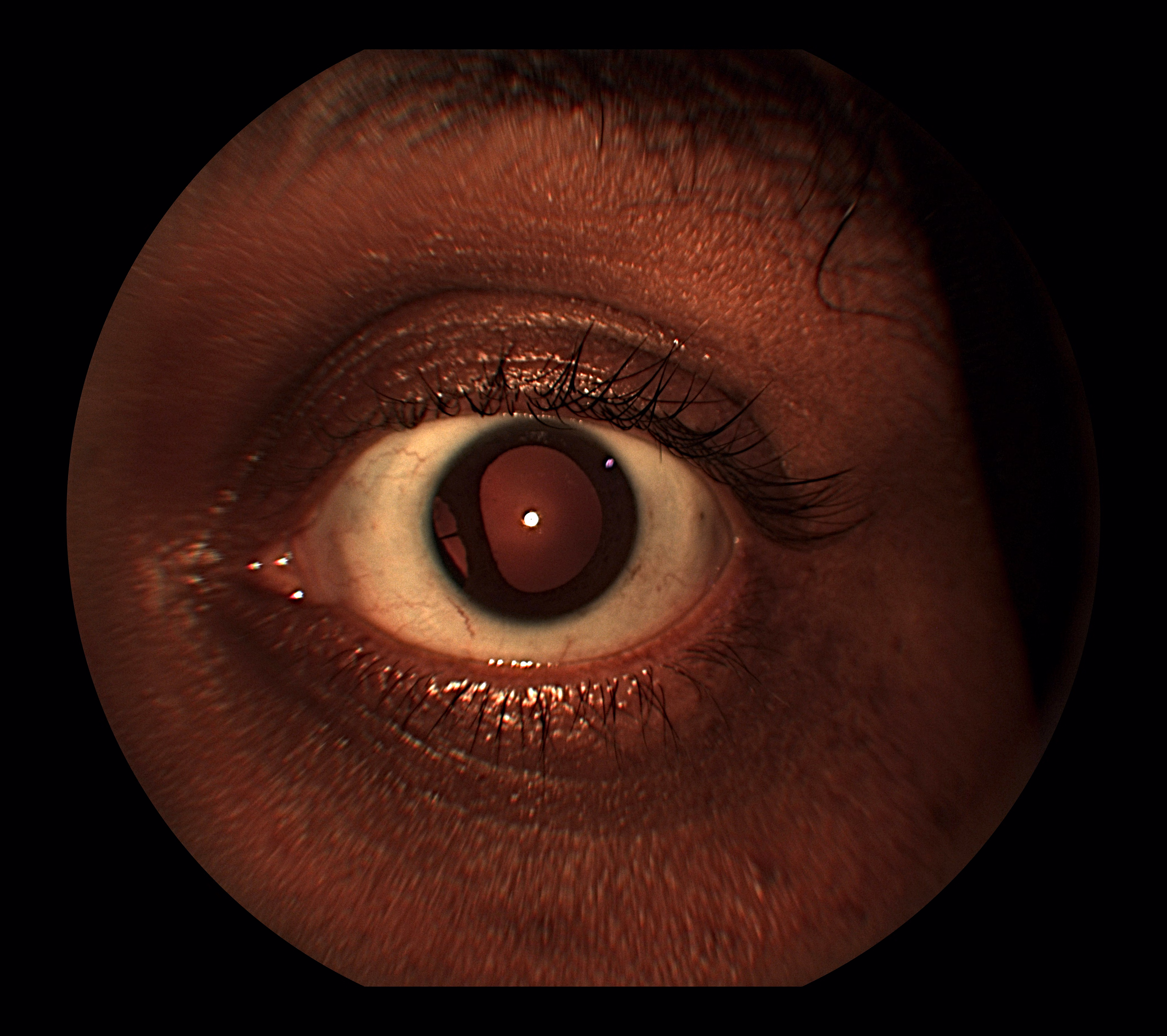
Go beyond retinal imaging
High-resolution external eye images allow for documentation of ocular surface and adnexal conditions.
QR Code led patient engagement
Engage, empower & educate patients via an interactive patient report that unlocks critical information on chronic retinal diseases.
Specifications
Resolution - 20.2 Megapixel
Fixation Targets - 4 Internal (Disc, Macula, Superior, Inferior) and External
Image Modes - Color, Red Free, Anterior Diffused and Posterior Reflex Free Imaging
Minimum Pupil Size - 5mm
Capture Mode Posterior - Auto-Focus and Auto-Capture
Field of view in single shot - Typically 58°, Up to 60°
Montage Options - 2 Images, 4 Images, 87° x 58° (2-field), 87° x 116° (4-field)
Contact us for
Powered by a 20 MP image sensor and a patented optical design
Wider Field enabling detection of peripheral disease
7-field ETDRS recommends a 75-degree FOV for early detection of Diabetic Retinopathy. With just two clicks, Pristine 5.0 offers upto 87° X 58° FOV*# and up to 87° X 116° FOV*# with four clicks.
*Visual Angle
#Horizontal x Vertical
Precision in Clinical Decision
Experience everyday precision with sharp detailing in images that identifies even hard-to-detect pathologies and tertiary blood vessels.
Go beyond retinal imaging
High-resolution external eye images allow for documentation of ocular surface and adnexal conditions.
QR Code led patient engagement
Engage, empower & educate patients via an interactive patient report that unlocks critical information on chronic retinal diseases.
Specifications
Resolution - 20.2 Megapixel
Fixation Targets - 4 Internal (Disc, Macula, Superior, Inferior) and External
Image Modes - Color, Red Free, Anterior Diffused and Posterior Reflex Free Imaging
Minimum Pupil Size - 5mm
Capture Mode Posterior - Auto-Focus and Auto-Capture
Field of view in single shot - Typically 58°, Up to 60°
Montage Options - 2 Images, 4 Images, 87° x 58° (2-field), 87° x 116° (4-field)
| Established in: | 2021 |
| Bussiness Type: | Partnership |
| Bussiness Type: | Merchant |
| MSME: | Yes |
| Countries exporting to: | Nepal, United States, Bangladesh, Sri Lanka, South Africa, Nigeria |
| Export Turnover (3 years): | US$10,000 to US$50,000 |
| Certifications: | UDYAM |
| Countries would like to export to: | Argentina, Australia, Austria, Azerbaijan, Bangladesh, Belgium, Brazil, Cambodia, Canada, China, Colombia, Cuba, Czech Republic, Denmark, Dominican Republic, Egypt, Ethiopia, Finland, France, Georgia, Germany, Greece, Hong Kong S.A.R., Hungary, Iceland, India, Indonesia, Iraq, Ireland, Israel, Italy, Japan, Kenya, Korea South, Kuwait, Lebanon, Luxembourg, Macedonia, Madagascar, Malaysia, Maldives, Mali, Mauritius, Mexico, Monaco, Mozambique, Myanmar, Namibia, Nepal, Netherlands Antilles, Netherlands The, New Zealand, Nigeria, Norway, Oman, Panama, Papua new Guinea, Peru, Philippines, Poland, Portugal, Puerto Rico, Qatar, Romania, Russia, Saudi Arabia, Singapore, South Africa, Spain, Sri Lanka, Sweden, Switzerland, Taiwan, Tanzania, Thailand, Turkey, Ukraine, United Arab Emirates, United Kingdom, United States, Uzbekistan, Vietnam, Yemen, Yugoslavia, Zambia, Zimbabwe |
| No of employees: | 1-10 |


